This story originally appeared in the Winter 2025 edition of On the Cusp®, Stony Brook School of Dental Medicine's semiannual magazine.
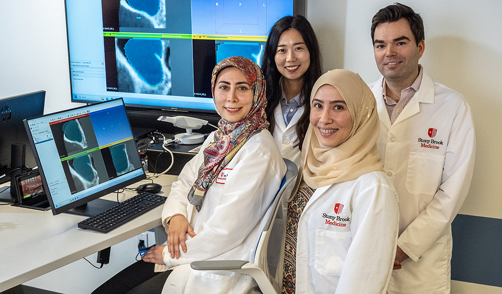
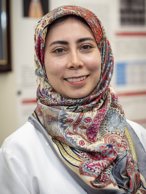
Mina Mahdian, DDS, MDSc, is helping to lay the foundation for artificial intelligence in dentistry — step by step, study by study.
As the world of AI expands and accelerates at a blistering pace, the director of the Advanced Education Program in Oral and Maxillofacial Radiology at Stony Brook School of Dental Medicine understands that the precision, diligence and patience that she and her colleagues demonstrate now, are crucial to supporting all the developments that follow.
Together with the residents she oversees, fellow researchers across the Stony Brook University campus, and scientists around the world, Mahdian has brought about significant progress in the various applications of AI in diagnostic imaging.
“It is gradually making its way into healthcare systems worldwide,” said Mahdian, an associate professor in Stony Brook School of Dental Medicine’s Department of Prosthodontics and Digital Technology. “It is definitely growing and is going to become a major part of dentistry, specifically precision dentistry. I can’t tell you how soon that will happen, but it is going in that direction.”
Enhancing Efficiency
Mahdian is a member of the Global Initiative on Artificial Intelligence for Health, Dental Diagnostics and Digital Dentistry, a mission launched by the World Health Organization (WHO), International Telecommunication Union (ITU) and World Intellectual Property Organization (WIPO). Its aim is to improve healthcare worldwide using AI and promote the exchange of knowledge and the development of technical standards for the use of AI in the healthcare sector.
She views AI as a complementary tool, inspired by human intelligence, created by humans for humans to use. It has shown early signs of revolutionizing industries like manufacturing, finance and healthcare, particularly with her field — imaging and diagnostics. Like with many AI tools used to perform mundane tasks, dentists can free up valuable time with finely tuned AI automation — when the time is right.
“Clinicians sometimes need to perform quantitative assessment of structures to evaluate anatomical changes or progression of disease. If someone wanted to manually delineate the anatomical structure or the pathology for quantitative analysis in a volumetric study, it might take them up to 30 minutes,” she said. “With one click on an appropriately trained AI tool, the same result can be achieved in less than a minute.”
Multiply that by the thousands of scans being done annually and productivity and efficiency grow exponentially. AI development could prove especially beneficial for dentists practicing in Healthcare Professional Shortage Areas (HPSAs) with limited resources and access to specialists in their area. By leveraging reliable AI tools to assist with certain diagnostic and treatment planning tasks at an acceptable level of accuracy, Mahdian said, they can see more patients and perform more procedures.
She began her own journey in AI in 2017 as a junior faculty, collaborating with a colleague on optical coherence tomography (OCT), using light to stage incipient versus advanced carious lesions based on optical properties of the tissues. The advantage of this technology over conventional radiography is that it uses non-ionizing radiation. However, due to the nature of these images, they discovered that it may be difficult for dentists to interpret these images and distinguish carious and non-carious lesions.
 “We then worked to develop a deep learning algorithm to decode these images based on the signal intensity values of the OCT images so dentists can differentiate carious and non-carious lesions,” said Mahdian, whose work was funded by an American Academy of Oral and Maxillofacial Radiology (AAOMR) XDR grant.
Mahdian has directed Stony Brook’s OMFR residency program since its launch in 2017, and in a short time, it has made an impact in the field of radiology. The program’s first-ever resident, Jalal Bukhari, DDS, received the AAOMR’s Howard R. Raper Award for excellence in radiology and his commitment to a career in academia. That same year, Maryam Ajami, DDS, earned the organization’s Albert G. Richards Graduate Student Research Grant for developing an AI model using cone beam computed tomography (CBCT) data to detect vascular calcifications, incidents that are often missed by non-radiologists.
“A lot of times, these calcified plaques in arteries can become clinically significant, especially if the patient is not aware of their existence, and may lead to reduced blood flow to the brain, and potentially cerebrovascular incidents (stroke),” Mahdian said.
Then, in a collaborative effort with Renaissance School of Medicine Department of Radiology faculty, Chuan Huang, PhD, Lina Albitar, DDS, MS ’22, OMFR ’22, trained a CNN-based AI model to identify missed second mesiobuccal canals in endodontically treated maxillary molars. These canals are often difficult to detect by human eye and may be a cause of failure of endodontic treatment. Dr. Albitar’s research earned the Best Resident Poster in the Research Category at the 2021 annual AAOMR meeting.
An even more recent endeavor extends into the field of radiomics where automated feature extraction is utilized to predict disease. Together with Prateek Prasanna, MS, PhD, in Stony Brook University’s Department of Bioinformatics and Haibin Ling, MS, PhD, in its Department of Computer Science, Mahdian applied radiomics to characterize vascular calcifications in CBCT images to determine a patient’s risk for cardiovascular disease. CBCT scans of the maxillofacial complex often include incidental findings beyond the teeth and the jaws, that may require medical follow-up, one such example being vascular calcifications. The patient may be unaware of the presence of these calcifications and the dental provider may be the first to identify these potentially significant findings and refer for medical evaluation. The trio received seed grant funding from the university’s Office of the Vice President of Research earlier this year.
“I am quite excited about this particular project, as this is the first study to apply radiomics to characterize vascular calcifications in CBCT to predict cardiovascular disease,” Mahdian said. “The majority of current AI research is focused on common dental pathologies, whereas this project, signifies the role of AI in assisting dental providers with predicting the risk of cardiovascular incidents based on dental CBCTs and making proper and timely referral.”
The Road Ahead
Challenges remain in the development of reliable AI tools. One of them is data, of which scientists need scores of in order to train models to perform efficiently and, even more importantly, produce accurate results. Lack of large, standardized datasets, that are of high-quality and representative of diverse populations poses a challenge to the development of reliable AI tools in dentistry. Furthermore, powerful AI servers are needed, to process these large quantities of data.
And, Mahdian emphasized, dental AI models must be validated by expert clinicians, and for advanced applications, oral and maxillofacial radiologists, of which there are only about 400 in the United States. Stony Brook is one of just 10 dental schools in North America that has a CODA-accredited oral and maxillofacial radiology residency program, and the only such program in New York State.
“There is a bottleneck right now for the development and validation of these tools,” she said.
Stony Brook as a whole is on the front lines of AI innovation. In September, it launched the AI Innovation Institute (AI3), which was created to coordinate, organize and advance AI innovation and education across campus. According to University Provost Carl Lejuez, PhD, the AI3 will “[catalyze] core AI research, curriculum innovation, and societal change in the ever-evolving landscape of knowledge work.”
Mahdian has mentored numerous students and residents in AI-related projects. Among them is Sonya Movassaghi, DMD, who enrolled in Stony Brook’s OMFR residency program in 2023 after spending eight years as a general dentist. Under Mahdian, and in collaboration with the Stony Brook Applied Mathematics Department, she has worked to develop an automated model to detect mucosal thickening within the maxillary sinus in CBCT images, measuring the volume of disease as well as the surrounding air and anatomical structures.
“By feeding images of both diseased and healthy sinuses to the AI algorithm, we can teach it what to look for and test its effectiveness in distinguishing mucosal thickening from healthy air, bone structure and the nasal cavity,” said Movassaghi. “Working on this project has opened my eyes to the power of AI and how rapidly this field is advancing and changing.”
However, its growth, between detection, efficiency and, in the end, patient care, is only as good as the foundation underneath it. That’s why Mahdian and her team are taking the necessary measures now so that those in the future, wherever they are practicing, can stand on their shoulders.
“A lot of people are concerned that AI is going to replace practitioners; I don’t think that will ever happen,” she said. “AI lacks consciousness, creativity, intuition and clinical judgment. It makes decisions mathematically. You ultimately need to have a person in charge and making decisions. AI has the potential to be a great complementary tool. I think we should all be open to testing it, seeing how it works, and trying to improve it, because it’s only going to help.”