|
21 year old female referred for endodontic therapy of tooth #3.
The patient is asymptomatic and states, "My dentist sent me her for a root canal."
The patient denies any significant medical history.
The patient has received routine dental treatment for the last 15 years.
She is currently under the care of a general practitioner. Tooth #3 was treated for deep decay resulting in a carious exposure. A temporary restoration was placed, and the patient was advised the tooth required endodontic therapy.
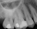
No swelling was noted extraorally or intraorally. Tooth #3 was asymptomatic. No mobility was noted. The tooth was not sensitive to percussion, and probing depths were 2 to 4 mm. A temporary filling was noted. Radiographic evaluation was within normal limits.
Pulpal: Irreversible Pulpitis secondary to caries
Periapical: Normal
Non-surgical root canal therapy #3.
Visit 1 — 1/11/01 
- Preoperative radiograph taken.
- Informed Consent gained.
- Topical, 36 mg lidocaine with 0.18mg epinephrine administered by buccal and palatal infiltrations.
- Rubber dam isolation.
- Tooth taken out of occlusion.
- Temporary restoration removed, revealing pulpal exposure of mesiobuccal pulp horn.
- Access gained.
- Working lengths obtained radiographically.
- Full instrumentation using Profile NiTi rotary files, NaOCl, Glyde, and K files.
- Cotton placed in the chamber, and the tooth sealed with Cavit.
Visit 2 — 1/17/01
- Topical, 36 mg lidocaine with 0.18mg epinephrine administered by buccal and palatal infiltrations.
- Rubber dam isolation.
- Cavit, cotton removed.
- Patentcy confirmed with 20 K file.
- NaOCl irrigation.
- Canals dried with paper points.
- Obturated all canals (gutta percha, Roth's sealer, warm vertical condensation).
- Cotton and Cavit placed.
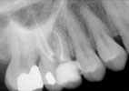
- Post-operative radiograph taken.
- Post-operative instructions given, and patient was referred to her general practitioner for the final restoration.
Visit 3 — 2/26/01
- Patient presents for emergency visit and complains, my dentist did a filling on the tooth behind the tooth you treated, and it hurts.
- Tooth #2 tender to percussion, thermal testing yields normal responses (compared to other teeth in quadrant) with no lingering sensations.
- Articulating paper reveals tooth #2 is in hyperocclusion.
- Occlusion on #2 was reduced.
- Patient advised to wait to determine if symptoms get better or worse, and follow up in 3 to 4 weeks.
Patient presents and states, "it's getting worse."
No swelling was noted extraorally or intraorally. Tooth #2 is tender to palpation, hypersensitive to cold, with lingering sensation for 15 seconds. Probing depths were 2 to 4 mm, no mobility was noted. An amalgam filling was noted which radiographically approximated the pulp.
Pulpal: Irreversible Pulpitis secondary to caries
Periapical: Acute Apical Periodontits
Non-surgical root canal therapy #2.
Visit 4 — 3/12/01 
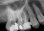
- Preoperative radiograph taken.
- Consult with patient's general practitioner to confirm treatment plan.
- Informed Consent gained.
- Topical, 72mg lidocaine with 0.36 mg epinephrine administered by buccal and palatal infiltrations.
- Rubber dam isolation.
- Tooth taken out of occlusion.
- Amalgam restoration removed, a pulpal exposure was noted on the floor of the preparation.
- Access gained.
- Pulpectomy performed.
- Irrigated with sodium hypochlorite
- Cotton placed in the chamber, and the tooth sealed with Cavit.
Visit 5 — 3/15/01
- Patient presents complaining of, "throbbing pain."
- Topical, 36 mg lidocaine with 0.18mg epinephrine administered by buccal and palatal infiltrations.
- Rubber dam isolation.
- Working lengths obtained radiographically.
- Full instrumentation using Profile NiTi rotary files, NaOCl, Glyde, and K files.
- Cotton placed in the chamber, and the tooth sealed with Cavit.
Visit 6 — 4/6/01 
- Patient presents asymptomatic.
- Topical, 36 mg lidocaine with 0.18mg epinephrine administered by buccal and palatal infiltrations.
- Rubber dam isolation.
- Cavit, cotton removed.
- Patency confirmed with 20 K file.
- NaOCl irrigation.
- Canals dried with paper points.
- Obturated all canals (gutta percha, Roth's sealer, lateral condensation).
- Cotton placed in the chamber, and the tooth sealed with Cavit.
- Post-operative radiograph taken.
- Post-operative instructions given, and patient was referred to her general practitioner for the final restoration.
This case was diagnosed and treated by:
Dr. Michael E. Newman, second year resident
Post-Graduate Endodontics
|


